
During the seminar, we showcase new features of TissUUmaps 3.1, such as:
TissUUmaps is designed to display and share spatially resolved data on top of a whole slide tissue image. Any kind of image and markers can be visualized and shared with anyone via web-browser without downloading the image data or installing any software.
You and your collaborators can also interact with the data. You can visualize spatial transcriptomics, in-situ sequencing, and data associated with cells, like morphology or cell types. All on top of the tissue to create some interesting tissue maps!
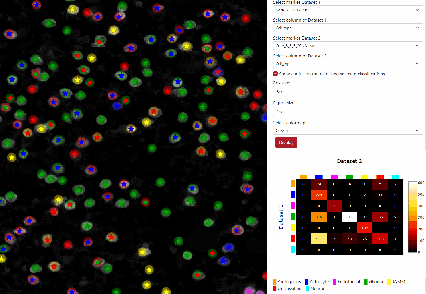
You can find a written tutorial how to use ClassV&QC plugin at this link.
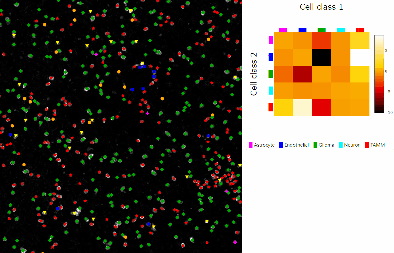
You can find a written tutorial how to use InteractionV&QC plugin at this link.
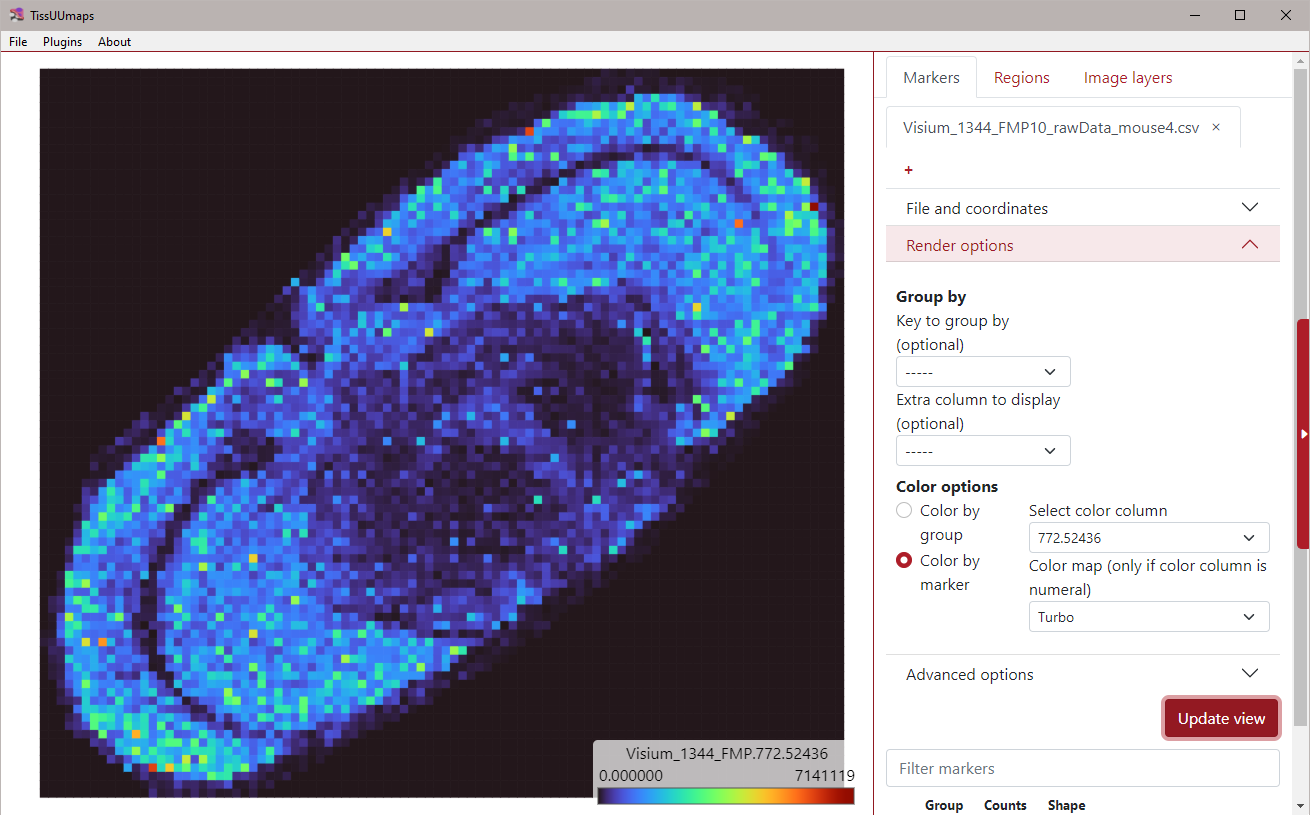
You can find a written tutorial about how to explore multidimensional data in TissUUmaps at this link.
This video is from the I2K 2022 (From Images to Knowledge) conference on May 10, 2022.
This video is from the BioImage Informatics webinar, where we present the standalone TissUUmaps 3.0.
In this video, we show feature of the TissUUmaps tool that is including feature space into the visualization of the tissue section overlaid by markers.
In this video, we show how to use TissUUmaps to visualize the outputs from two well known ST analysis frameworks: Giotto and Squidpy.
The original tutorial for analyzing 10X Visium (Giotto) can be found here, and the code for integration with TissUUmaps in giotto2tmap.py.
The original tutorial for Analyze Visium H&E data (Squidpy) can be found here, and the code for integration with TissUUmaps in squidpy2tmap.ipynb.
TissUUmaps allows to visualize convolutional neural network (CNN) on multiresolution images. In this tutorial, we show how to extract morphological features from any spatial transcriptomics (ST) dataset using any pretrained CNN. In the end, we will have a vector of features for every ST spot in the image, that we can then reduce to 3 dimensions and map back to the image as RGB colors.
You can find the Jupyter Notebook in: tissuumaps_cnn_example.ipynb .
This tutorial will show you how to integrate a code from Jupyter notebook with our interactive viewer TissUUmaps. For this example we use the method Spage2vec - unsupervised segmentation-free approach for decrypting the spatial transcriptomic heterogeneity of complex tissues at subcellular resolution.
You can find the Jupyter Notebook in: spage2vec_TM.ipynb .
In this tutorial we will look at how one can TissUUmaps to explore raw situ sequencing data. In this tutorial we use TissUUmaps windows desktop client together with a plugin called Spot Inspector.
See github.com/TissUUmaps/TissUUmaps for installation instructions.
In this video we show how to run Cellpose to segment cells in Napari, export the resulting images as a TissUUmaps project, and finally load the project in TissUUmaps web.
Note that the napari-tissuumaps plugin can export images, masks, but also point clouds and regions in vector formats.
More info at: github.com/TissUUmaps/napari-tissuumaps
This tutorial shows how to visualize starfish decoding experiments in TissUUmaps. It follows the oficial starfish Pixel-Based Decoding tutorial and then converts the outputs to a compatible format to the spot inspector plugin for quality control.
Find the JupyterNotebook at pixel_decoding_starfish.ipynb and the helper functions at starfish2tmap.py.
In this video we see what TissUUmaps can do and what data we can visualize and explore. TissUUmaps allows the display of tissue slide images and uses an overlay to display any sort of marker data on top. Be it spatially resolved gene expression, per cell data, or regions of interest.
Any image can be converted to a pyramidal image and point TissUUmaps to it so it can be displayed, it is not limited to tissue data but it is its main purpose. The images are not used for processing or extracting information only visualization. Since the images are displayed via the web, only 8-bit images can be displayed. If you have a different bit-depth you will need to convert it to 8-bits before using VIPS.
This video will focus on the gene expression data. A CSV file that contains either a barcode or a gene name or both, associated to an X,Y position in the image. More data can be associated to the coordinate, all of it is available in the TissUUmaps interface.
This video focuses on the creation and use of regions to explore data. Regions are a JSON file containing the polygons that describe the regions. These regions can be saved and imported.
Use the browser console and developer tools to create new functionality and new visualizations of your data. We offer a basic organization of it with our API.